


Splinting for Ligament Sprains: "Why Is It Necessary?" The 3 Anatomical Phases of Healing
Ligaments are specialized tissues composed primarily of collagen fibers. When these fibers are torn due to a sprain, the human body initiates a precise biological reconstruction process: Inflammatory (Days 1–5) → Proliferative (3 days–3 weeks) → Remodeling (3 weeks–months). In each phase, a splint plays a critical role; improper stabilization is a leading cause of chronic instability.
1. The 3 Phases of Anatomical Recovery: The Role of Splinting
① Inflammatory Phase (Days 1 – 5)
Immediately after injury, the body enters a state of 'emergency.' Blood vessels dilate, and leukocytes and macrophages flock to the site. Their mission is to clear debris and dead cells, preparing the "construction site" for repair.
- Cellular Response: Vasodilation → Recruitment of leukocytes/macrophages → Debridement of the injured site.
- Role of the Splint: Micro-movements during this stage can cause further bleeding and pain. The splint immobilizes the joint to prevent excessive inflammation and serves as a physical barrier to minimize edema (swelling).
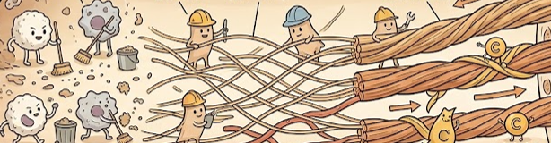
As shown above, a splint effectively blocks micro-movements, allowing the natural healing process to begin without interruption.
② Proliferative Phase (3 Days – 3 Weeks)
This is when new tissue generation begins. Fibroblasts proliferate and begin laying down Type III Collagen, which acts as a temporary framework. New micro-vessels also form to supply oxygen and essential nutrients.
- Cellular Response: Fibroblast proliferation → Type III collagen framework deposition → Neovascularization (new blood vessel growth).
- Role of the Splint: It ensures that the ligament fibers do not heal in a "slack" or lengthened position by maintaining correct anatomical alignment. Removing a splint too early during this phase can result in a loose ligament, leading to permanent instability.
Type III collagen provides the initial bridge but lacks the strength of mature tissue. The splint guarantees precise alignment during this delicate transition.
③ Remodeling Phase (3 Weeks – Several Months)
This is the "upgrade" stage. The weaker Type III collagen is replaced by the much stronger and more durable Type I Collagen. Disorganized fibers begin to align along the lines of physical stress, restoring the ligament's original strength.
- Cellular Response: Transition from Type III to Type I collagen → Fiber alignment → Restoration of tensile strength.
- Role of the Splint: It acts as a bridge from total immobilization to gradual mobilization, protecting the tissue until it can withstand external loads.
2. Nutritional Allies: Supplying the "Construction Site"
For the biological repair under the splint to succeed, a steady supply of "raw materials" is essential. Collagen synthesis, in particular, is highly dependent on specific nutrients.
- Amino Acids (Proteins): The basic building blocks of collagen. Consuming high-quality proteins rich in Proline and Glycine supports fibroblast activity.
- Vitamin C: Acts as a "cross-linking agent" that knits collagen molecules together. Without enough Vitamin C, the resulting ligament will have significantly lower tensile strength.
- Omega-3 & Antioxidants: Help regulate the initial inflammatory response and reduce oxidative stress, accelerating cellular regeneration.
- Zinc: Critical for cell division and protein synthesis, promoting tissue growth during the proliferative phase.
3. The Manufacturer’s Perspective: Why Quality Matters
Understanding the anatomical healing process highlights why the quality of a splint is paramount. Substandard splints can hinder recovery and cause secondary complications.
- Uniform Pressure Distribution: Uneven pressure can obstruct blood flow, cutting off the oxygen supply needed for tissue proliferation.
- Ventilation: Increased cellular metabolism generates heat and sweat. If these aren't managed, skin complications arise, diverting the body's immune resources away from the ligament.
- Strength vs. Weight: A splint must be light enough for the patient to wear consistently through the remodeling phase while providing uncompromised stability.
References
[Anatomical & Physiological Basis]
- Frank, C. B. (2004). Ligament injury, healing and repair. Journal of Musculoskeletal and Neuronal Interactions.
- Akeson, W. H., et al. (1984). The connective tissue response to immobility: An overview. Connective Tissue Research.
- Guyton and Hall (2020). Textbook of Medical Physiology, 14th Edition. Elsevier.
[Nutritional Science]
- Papadopoulou, S. K. (2020). Rehabilitation Nutrition for Injury Recovery of Athletes. Nutrients.
- Shaw, G., et al. (2017). Vitamin C-enriched gelatin supplementation before intermittent activity augments collagen synthesis. Am J Clin Nutr.
⚠️ Disclaimer: This guide is for informational purposes only. Every patient's injury and recovery speed is unique. Always follow the diagnosis and prescription of an orthopedic or rehabilitation specialist. Improper use or adjustment of a splint may lead to circulation issues or nerve damage.

