



Finger Splint Muscle Support and Anatomical Key Points
Key Supporting Muscles
The flexor digitorum profundus & superficialis¹ control finger flexion, particularly stabilizing the distal interphalangeal joint (DIP joint)² and proximal interphalangeal joint (PIP joint)³. Finger splints prevent excessive strain and re-injury to these muscles.
The extensor digitorum⁴ regulates finger extension, maintaining alignment of the metacarpophalangeal joint (MCP joint)⁵ and PIP joint³. The splint protects the functional positioning of this muscle.
Intrinsic hand muscles including the lumbricales⁶ and interossei⁷ control fine finger movements and abduction/adduction. Finger splints maintain balance in these muscles.
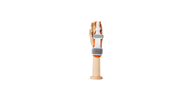
Nerve Protection and Precautions
The median nerve and ulnar nerve govern finger sensation and motor function. Finger splint application must prevent nerve compression that could cause numbness, sensory loss, or motor weakness.
Thumb and index finger areas require median nerve protection, while ring and little finger areas need ulnar nerve safeguarding. Sensory testing of patients is essential.
Application Precautions
Finger splints are typically positioned with the PIP joint³ at 30-40° flexion and DIP joint² at 0-15° flexion, requiring customized application based on each finger's anatomy.
Functional immobilization rather than complete fixation is the principle, with periodic active motion exercises needed during long-term wear to prevent muscle atrophy and joint stiffness.
Adequate padding, breathable materials, and regular skin checks prevent skin issues, swelling, and circulatory problems.
Footnotes
References
⚠️ This information is general guidance only. Individual patient conditions may vary, so medical professional evaluation and consultation are essential.

